< | up
Small Blue Round Cell Tumor Enigma (18832)
Small Blue Round Cell Tumor Enigmanew
Subtitle: A10-3
Type:
SOFT
Sender:
ugnius
2010-02-06 13:33
INCTR - Lithuanian Pathology online
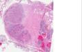
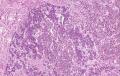
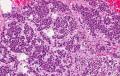
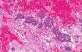
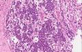
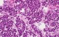
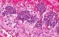
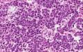
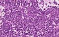
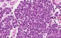
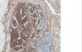
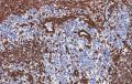
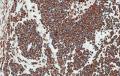
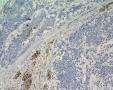
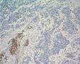
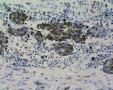
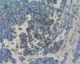
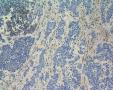
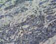
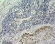
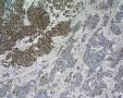
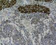
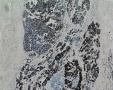
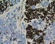
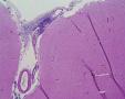
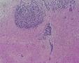
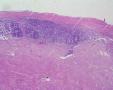
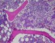
3 years girl was hospitalized due to lower limb paresis. on CT scan small tumor nidus was found in retroperitioneal left lumbar area with spread to spindal cord, mts to the cerebral meninges. The patient was deceased soon due to difusse cerebromalacia/liquor drainage blockage. Autopsy shows the same tumoral spread (difficult visualisation due to small mass), prominent neuro- tropism and distant mts to bone marrow and CNS coverings.  
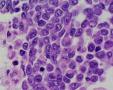
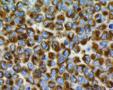
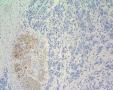
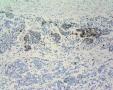
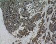
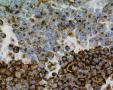
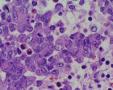
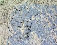
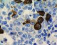
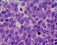
Clinically and at first glance histologically the idea of neuroblastoma possibility was evolved. Any clinical/molecular/biochemic analyses regarding neuroblastoma version were done due to rapid course of the disease.  
Immunophenotype: Vimentin+ 20%; PanCK+ 10%, Cam 5.2+ 5%; NB84+ 60%; EMA+ 70%; ChromograninA/Synaptophysin(-); S100+ 10%; Desmin/MyoD1 (-), CD99(-)(single focus <5%+), CD56(-), NSE+ 10% (patchy), CD68/MelanA/HMB45(-), Hepa/CD57/CD34/NFP/GFAP(-), CyclinD1(-), CD20/CD30/CD3/LCA(-); p53+ 10%. Ki67 prolif. activity ~40%.  
PROPOSAL: Small blue round cell tumor with rhabdoid features, maybe extrarenal rhabdoid tumor (paraspinal) vs epithelioid MPNST (exceptional in this age group) with spread to spinal cord and mts.
Annotations » Add comment (Login)
frankst
2010-02-08 10:39
To me, (cyto)histomorphological aspect and immunoprofile as well as clinical presentation (incl. leptomeningeal dissemination) raise the possibility of an atypical teratoid/rhabdoid tumor (AT/RT). To prove/disprove this diagnosis, IHC for INI-1 (which should show loss of nuclear expression in tumor cells, while being retained in normal cell nuclei (e.g. endothelial cells) seems required. AT/RTs can occur as part of the hereditary rhabdoid tumor predisposition syndrome.
ugnius
2010-02-08 10:52
Thank you for prompt reponse. Unfortunatelly I have not INI in my armentarium up to date. AT/RT was/is in the differential, but I have no "teratoid" components here. Really "rhabdoid" features present in minority of the cells. Predominant population represents something like "undifferentiated" cells... I will proceed with left IH panel, but hopefully nothing new will appear... Thank you one more time.
ugnius
2010-02-09 16:44
The last negative IH arrived: included into general IH description.
frankst
2010-02-10 09:25
For me, AT/RT continues to stay on the top of the list of potential diff. diagnoses. Please extend IHC by smooth muscle actin and p53 (the latter to exclude (?) MPNST, as the vast majority of them should be positive). I could also offer to perform IHC for INI-1, if you could send me a few unstained slides (Institute of Pathology, University of Basel, Switzerland).
ugnius
2010-02-10 09:38
Thank you a lot. Asm Actin in general negative (single cells only +: I will post it in seconds). I will check p53 additionally. Several unstained slides will be sent to you soon. Thank you for this unique possibility. Maybe it would be a possibility for INI1 molecular test? May I append parafin block for that?
frankst
2010-02-10 10:00
[comment sent by email]
No, we`d just do INI-1 IHC. So unstained sections would suffice.
 
Please send a generous number in case INI-1 is retained.....
 
 
ugnius
2010-02-10 10:24
Thanx. Do my best.
ugnius
2010-02-11 14:41
The last IH: LCA/CD20/CD3/CD30(-). p53+ 10% (photo apended). The slides are sent to Basel.
ugnius
2010-02-20 07:25
Ewing Sa (FISH) and DSRCT (PCR) molec's negative.
frankst
2010-03-01 11:20
[comment sent by email]
So far, I have not received any sections. Would refrain from
 
initaiting additional diagnostic tests, as long as the INI-1 status
 
is unclear.
 
 
ugnius
2010-03-01 11:43
Dear Stephan Frank, the slides were sent to your Institute in 12 February (2 weeks ago, avia mail). I'm worried about that.
frankst
2010-03-04 09:37
Just received the sections (in good shape; March 4th). Will proceed with IHC for INI-1 as arranged.
ugnius
2010-03-04 09:41
I'm really happy to hear it. Waiting NEAGATVITY:)
frankst
2010-03-08 15:10
[comment sent by email]
It`s negative (and I stick with my initial diagnosis).
 
 
ugnius
2010-03-08 15:30
Thank you a lot. What argues you propose CONTRA extrarenal rhabdoid tumor entity and PRO AT/RT? Really it's my first case in my 15 year practice.
frankst
2010-03-09 11:20
[comment sent by email]
Good question. Considering that about 20% of AT/RTs present with CSF
 
dissemination already at the time of diagnosis (as was the case
 
here), I`d still entertain an AT/RT. In addition, I don`t know
 
whether extrarenal rhabdoid tumors express S100 (as in your case). If
 
that is the case, one might of course also very well regard this
 
tumor as extrarenal rhabdoid tumor, and you are absolutely right with
 
your comment; in addition, the retroperitoneal manifestation should
 
then be interpreted as extrarenal RT. Along the same line I have to
 
admit, that as "pure" neuropathologist, I don`t have a lot of
 
experience with that latter entity. In the end, it`s an highly
 
academic question, I suppose.
 
 
ugnius
2010-03-09 11:42
Thank you for all done. Maybe in future it would be of interest to publish the case report with your kind participation?
frankst
2010-05-04 17:54
[comment sent by email]
I am happy to assist. At this point, as this case is solved, we
 
should probably shift conversation to normal Email (frankst@uhbs.ch).
 
Please send me a first mail, as I don`t have your correct address.
 
 
» Add comment (Login)
Last modified: 2011-07-05 10:42:40