< | up | >
Reactive lymphadenopathy, dermatopathic lymphadenitis (4259)
Reactive lymphadenopathy, dermatopathic lymphadenitisin review
paantraštė: Bilateral inguinal lymphadenopathy in a 24-year-old man
rūšis:
inguinal lymph node
siuntėjas:
semir
2007-12-05 19:36
INCTR - EBMWG Hematopathology Online
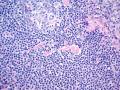
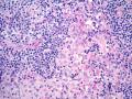
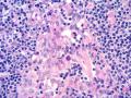
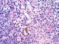
We present here a new case of the the enlarged inguinal lymph node, extirpated in a 24-year-old Bosnian male.  
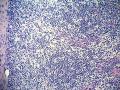
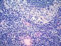
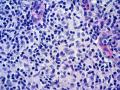
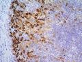
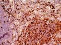
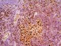
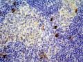
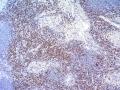
He was admitted to the Department of Infectious Diseases due to the fever and enlarged, painful inguinal lymphadenopathy follow by malaise and weight loss.  
WBC: Anemia and neutrophilia; CRP 184;Increased Alpha2 and Gamma globulins.  
Clinical examination revealed also hepatosplenomegaly and right axillary lymphadenopathy confirmed by ultrasonography. CT scan showed retroperitoneal lymphadenopathy.  
Microbiology: Negative for Toxoplasma, Cytomegalovirus, Herpes simplex, EBV, Mycoplasma pneumoniae, Brucella sp., Coxiella Burnetii. Serial analysis of his stool revealed no pathogenic microorganisms.  
He has been treated with antibiotics but without effective results.  
An inguinal lymph node, that measured 2.8x1.5x0.8 cm was received for histopathological examination.  
Histopathological examination showed generally preserved the structure of the lymph node. It also revaled dilated sinuses with histiocytes, lymphocytes and plasma cells. Focally, in subcortical areas sporadic histiocytes, lymphocytes, plasma cells and eosinophyls were noticed. In interfollicular areas some plasma cells and eosinophils were seen.  
Immunohistochemistry: CD1a(+), S100(+), CD68(+), CD3(-), CD20(-), CD30(-).  
Clinical diagnosis: No  
Histopathological diagnosis: Reactive lymph node?  
Please, give your opinions.
anotacija » pridėti komentarą (prisijungti)
Mueller-Hermelink
2007-12-05 20:09
Thank you for these nice photographs. This is clearly a reactive process of a so called nonspecific lymphadenitis showing features of sinus histiocytosis ( resorptive process ?) and dematopathic lymphadenopathy ( chronic inflammatory processes in the overlying or site dependent skin area) This explains the increase of CD 1a + dendritic and also probably Langerhans cells in the marginal sinus and T cell areas which are also S100 + . There is no suspicion of Langerhans cell histiocytosis!
franco
2007-12-05 20:43
I fully agree with Prof. Mueller-Hermelink diagnosis of reactive non-specific lymphadenopathy, prevalently sinusal. However, inguinal lymph nodes could not be representative of the process.
anpo
2007-12-08 16:43
I agree with Vito - one should probably proceed with examination of the axillary lymph node. This inguinal lymph node does not explain why the patient has hepatosplenomegaly.
torlakovic
2007-12-12 06:58
I also agree that morphologically and IHC show no alarming features and are quite fine for paracotical hyperplasia with Langerhans cells most compatible with dermatophatic lymphadenopathy. I also agree with suggestions that this in itself most of the time will be hard to use to justify hepatosplanomegaly, weight loss and right axillary lymphadenopathy quite far from draining areas of inquinal LNs. Let me ask about intracytoplasmic inclusions-like dots in HE image no. 3 in histiocytic cells - melanin or something else? I was also a bit disturbed with your CD3 - not enough cells to cover for all lymphoid cells in this area. You unfortunately did not show CD20 image.
hurwitz
2007-12-14 16:50
In full agree ment with my colleges, I woul like to conclude this case with the diagnosis:  
Lymphadenitis with prominent sinus histiocytosis, consisitent with dermatopathic lymphadenitis.  
No evidence of malignancy.  
Comment: this finding cannot explain neither the patients symptoms, nor the hepatosplenomegaly or axillary lymphadenopathy. Depending on the clinical evolution an extirpation of an axillary lymphnode or a liver biopsy might be necessary.  
Thanks to the contributor and the participants in the discussion.
semir
2008-01-18 22:46
Just added five figures of the axillary lymph node, recently exctirpated in the same patient.  
Please, give your opinion, based on the previous findings and these new microphotographs.
diane.c.farhi
2008-01-21 16:40
[comment sent by email]
I agree with the previous comments, and I think that the second lymph
 
node shows essentially the same features. I, too, am concerned that the
 
findings do not explain the systemic symptoms, the retroperitoneal
 
lymphadenopathy, hepatosplenomegaly, and peripheral blood findings. I
 
think that additional serologic studies for different organisms are
 
indicated, as well as tests for lupus and related autoimmune diseases
 
(although the histology doesn't suggest it). I don't see any evidence
 
of malignancy in the slides submitted.
 
hurwitz
2008-01-24 20:39
Thanks for adding the images of the axillary lymph node, which as stated by Diane shows essentially the same findings as described in the inguinal lymph nodes. There is no evidence of malignancy.  
In retrospect I am asking myself it has been justified to suggest the biopsy of the axillary lymph node. The answer is yes, because now the probability that the clinical symptoms might be caused by an overlooked LPD has been practically eliminated.
» pridėti komentarą (prisijungti)
Last modified: 2007-12-05 19:36:13