< | up | >
PTCL with granulomatous reaction (4346)
PTCL with granulomatous reactionclosed
paantraštė: B07-32414
rūšis:
lymphnode and skin
siuntėjas:
ugnius
2007-12-10 10:38
INCTR - EBMWG Hematopathology Online
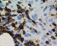
HISTORY: 79 yrs old female with uncertain diagnosis: 2 previous skin biopsies reveal granulomatous process, most probable "actinic granulomatous" (2007 August- September). Clin.: purple nodules and plagues on the face.  
Threpine biopsy: low quality, most probable reactive changes (no photographs).  
Lymph node biopsy was taken in November due to "disseminated lymphadenopathy": due to my "sympathy" to granulomatous processes the general IH panel was applied to this "sarcoid" like lymphadenitis with clusters of large cells with polylobated nuclei with a huge nucleoli in sinus and paracortical area, vague nodularity/septation: CD30+ CD43+ CD15- EMA- CD20- CD3- Bcl6-. GranzymB/ALK1/CD4/CD8/EBV LMP1 negative. The admixture of plasmacytes are present.  
 
PROPOSAL: cHL unclassified (probable NS 1?) with prominent granulomatous/sarcoid like reaction in LN.  
 
WORKING DIAGNOSIS: T lymphoma in the skin with the prominent granulomatous reaction and spread to LN.  
Thank you for participation.
anotacija » pridėti komentarą (prisijungti)
SergeyN
2007-12-10 11:35
It could be a lymphoma as well. A very interesting case, I think more stains will be necessary for a sure diagnosis: EBV, LCA (though it will probably be very difficult to interpret), CD79a, ALK, bcl2, some marker for cytotoxic phenotype. Maybe, CD5.  
 
Could you re-evaluate the skin biopsies?
ugnius
2007-12-10 12:59
Thanx. Please find above some negative stains. Some add are in progress.
ugnius
2007-12-10 13:01
I will apply to granulomatous skin lession similar IH panel. Visually HE without suspition of LPD.
hurwitz
2007-12-11 17:06
I agree with your diagnosis, Ugnius, I also favor the diagnosis of cHL, most probably nodular sclerosis, NS2, with prominent sarcoid like reaction. Arguments for this diagnosis are: Thickened lymphnode capsule and broad bands of collagen, seen on the panoramic images. Atypical cells, morphologically consistent with RS cells, CD30+, CD15 is negative, but this does not exclude the diagnosis. My impression is that one large atypical cell seen on image labelled "CD20 giant" is CD20+. This could be a hint to an EBV association. Did you ask for an EBV stain ?  
The occurrence of sarcoidosis or sarcoid like reactions in HL is a rare but well described phenomenom.  
Could you please submit some images of the skin biopsy and also of the bone marrow biopsy even if it is low quality. Just to get an impression.  
I am adding two ref. on this topic, the first an older one, very similar to the presented case. {PMID: 1420110} {PMID: 14499156}
tzankov
2007-12-12 09:07
have you thought on the possibility of a granulomatous cutaneous T-cell lymphoma with CD30+ anaplastic cells, e.g. granulomatous slack skin? partcularly CD15- CD43+ should let one think also of a T-cell lymphoma. I would extend the IHC pannel with CD5, CD4, CD8 and cytotixic markers (TIA1) and more precisely study the cutaneous infiltration to exclude T-cell lymphoma. does the patient has visible skin lesions? why was the skin biopsied?
ugnius
2007-12-12 09:20
Thank you. Please find skin changes- between confluent granulomas- scattered clusters of CD30+CD15-CD20-CD3- giant HL and RS like cells. So.... we proceed with cHL (NS) in LN and skin with prominent sarcoid/granulomatous reaction. I will take some photos regarding T NHL idea.
ugnius
2007-12-12 09:55
Really some atypical cells in the skin CD3 faintly +, but CD20-...
tzankov
2007-12-12 10:30
Though I have no experience with MUM1 in cutaneous T-cell lymphomas, I feel that the newly added pictures from the skin lesions should let one intensively think of a T-cell lymphoma, albeit skin infiltration by Hodgki lymphoma is extremely rare.
ugnius
2007-12-12 10:33
Scattered granzymB/Perforin+ cells are present betwen granulomas in LN. T population CD4+ >CD8+. CD2, CD5, CD7 in progress but I'm in doubt about informativity of evaluation of still scant population...
hurwitz
2007-12-13 17:18
Thanks Ugnius for the added images of the skin biopsy. Now we are facing a totally different situation. The skin infiltrates seem to be very similar, if not identical to the morphology seen on the lymph node, which brings us to consider a primary T-cell lymphoma, as the preferred diagnosis in particular granulomatous form of mycosis fungoides/ granulomatous slack skin as mentioned before by Dr.Tzankov. You mentioned two previous skin biopsies. Could you please add images of the other skin biopsy as well, and if possible also images of the clinical appearance of the lesions?  
I will try to ask a dermatopahologist to see the case.  
Regarding MUM1 expression, according to Tsuboi K et al,leukemia 2000,14(3)449-56 MUM1 staining was found in a large number of T-cell lymphomas including ALC, and in the majority of HL.  
ugnius
2007-12-13 20:46
Thanx. but really giant cell- single argue pro-tumor diagnosis; They are CD3-. The rest of T population is too scant and seems to be reactive/normal. I've recomended to check TCR arrang.
Mueller-Hermelink
2007-12-13 23:38
I am really a bit late in the discussion and agree with most of the comments . My primary feeling was PTCL because of these atypical clear cells . The large cells have to be looked at separately : are they HD-like cells ? maybe occuring as a superimposed EBV- driven proliferationor are they really T cells of an ALCL-like phenotype? We need some more immunophenotyping : Pax5 , LMP1 or EBER, Perforin TCR - rearrangement . This case can be solved !
schulze
2007-12-17 17:23
I agree with Dr. Tzankov and favor the diagnosis of specific skin infiltrate of granulomatous T-cell lymphoma with CD30+ anaplastic cells. Lymph node involvement speaks in favor of secondary skin involvement, whereas the lack of CD15 expression speaks against Hodgkin lymphoma.
ugnius
2007-12-20 14:42
Please find some add IH photos about T population CD4+: periadnexal, intersticial, perigranulomatous and some intraepidermal small cells with irregular nuclei. Maybe "Granulomatous" mycosis fungoides variant is most appropriate FINAL? Thank you.
SergeyN
2007-12-21 15:21
If I am not mistaken, "granulomatous slack skin" as a MF variant has a quite different clinical presentation. In addition, there should be destruction of elastic fibers.  
 
Skin T-cell LPD's could be accompanied by giant cell granolomas, the feature is hardly specific.  
 
tzankov
2007-12-21 15:34
In the presence of lymph nodes iwth such huge infiltration, I would primary diagnose a PTCL with (secondary) skin involvement, rather than a grnaulomatous MF.
ugnius
2007-12-21 15:51
Thanx. Discussing about PRIMARY SKIN T LYMPHOMAS: I mean the possibility mycosis (with unusual histo "granulomatous" appearance). The "cutis laxa" for sure is not present. If NOT- what entity from EORTC list you prefer else?  
If we will decide to call it peripheral T lymphoma, NOS with skin and LN damage- maybe it will not change the therapy scheme. Really all I can do- to check TCR. CLINICALLY?HISTORICALLY the first sign was progressing facial skin eruption. LN entrapment was secondary. But its not very strong argue for sure. The main problem is MISSED skin LPD at first biopsies...
ugnius
2007-12-21 20:41
"FULL" T panel IH in the skin: CD2+ like CD3+; CD5+ 60%; CD2+ 80%; CD7(-)(sic), CD56/CD57(-), GranzymB/Perforin(-).
hurwitz
2007-12-27 16:56
I would like to conclude this case according to the prevalent opinion:  
 
Peripheral T-cell lymphoma with prominent granulomatous reaction, involving lymph nodes and skin.  
 
Thanks to the submitter for this fascinating case, and thanks for all the interesting comments.  
» pridėti komentarą (prisijungti)
Last modified: 2007-12-10 10:38:07