< | up
Aspirate LT: quality control 1 (6526)
Aspirate LT: quality control 1new
Subtitle: B08-29685
Type:
Sender:
ugnius
2008-11-06 09:40
INCTR - Hematopathology teaching group
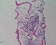
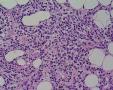
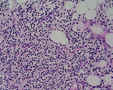
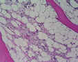
55 yrs male with threpine biopsy and aspirate.  
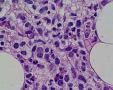
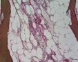
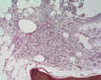
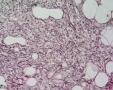
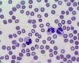
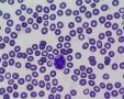
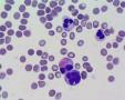
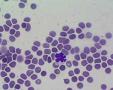
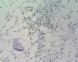
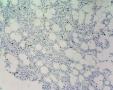
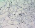
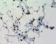
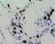
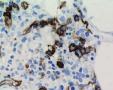
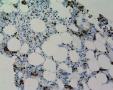
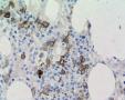
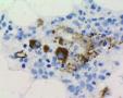
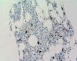
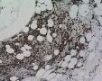
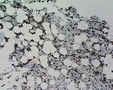
Diagnosis: aplastic anemia.  
History: 1 day patient from foreign country is under evaluation for BM transplant.  
Threpine hypocellular with fibrosis, arhitectural distortion, lymphoid agregattes and Fe deposition (MDS like picture). Only single micromegakaryocytes. Scattered NASDE+ granulopoietic cells, single erythroid cells, multiple eosinophils, maybe mastocytes are present. Some NASDE(-) precursor cells with "blastic" morphology. Blood test is attached.  
Smear: Photos are made with standard 60x lens. In case I have 100x oil in reserve:) An aspirate seems to be low cellular, foccaly overcrowded by erythrocytes.  
 
IH (hot spots): CD34/CD117+ precursors with slight clustering up to 20%; CD117+ mastocytes; Lymphos: CD3+ >> CD20+ (single); Single CD138+ plasmacytes; CD34+ neovascularity is prominent. vWF+ single micromegakaryocytes.  
 
WORKING: Architectural distortion with excess CD34+/CD17+ precursors up to 20% (scant hematopoietic population at all), III-IV fibrosis and prominent Fe deposition: high grade MDS is suspected. The use of CSF must be excluded.  
 
Please comment all diagnostic things, especially techno aspects and cytology.  
Thank you a lot.
Annotations » Add comment (Login)
SergeyN
2008-11-06 11:38
Dear Ugnius,  
 
you really need oil to evaluate the cytology quality. At this magnification the nuclear structure cannot be discerned, and it is difficult to say if it is a staining artefact.  
 
Color balance is a bit bluish, but it is not crucial.
diane.c.farhi
2008-11-06 19:18
I think the slide quality, both gross and microscopic is quite good. Lab values show monocytosis and normocytic normochromic anemia. I take it that the smears are of aspirate; they look hemodilute. One or two odd neutrophils are found, which are consistent with myelodysplastic change; this is OK for hairy cell leukemia (read on). ***For future reference, see if you can get the clinicians to do touch preps on all cores; these are invaluable when the marrow yields a dry tap, as in this case with fibrosis.*** The core biopsy shows thin bone with increased osteoclastic activity, typical of a metabolic etiology (this includes lots of syndromes, including B cell disease). The cellularity is patchy, with areas of hypoplasia. The more cellular areas show a fairly uniform population of small lymphoid cells surrounded by clear cytoplasm with sharp cytoplasmic borders. A close-up view shows elongated to indented or kidney-shaped nuclei. Fibrosis is marked. In summary, I feel the findings are typical of hairy cell leukemia. If you have a TRAP stain, you can try that on the smears. Also, I suggest going back to the peripheral blood are seeing if you can find hairy cells.
ugnius
2008-11-06 19:22
Thank you. I will proceed with IH later and append other cases with the smears for diagnostic/techno quality control:)
ugnius
2008-11-06 19:27
One more question: how to prevent hemodilution and get more concentrated material?
diane.c.farhi
2008-11-06 19:39
If the dry tap is due to locating the biopsy needle in cortical bone rather than medullary bone (usually easy to tell in the gross core specimen), it can be corrected by re-inserting the needle deeper into the bone marrow. If the dry tap is due to marrow necrosis or marrow replacement by fat, bone, fibrous tissue, and/or an abnormal infiltrate (as in your case), then there is no way to avoid it. The liquid aspirate can be concentrated by centrifuging the specimen, then aspirating the resulting buffy coat and a little blood, and using that to make slides. It is always a good idea to make several touch preps of the core to help in these circumstances. This must be done within a minute or two at the bedside. The first touch preps may be too bloody, so several slides should be made, until cells stop coming off the core.
ugnius
2008-11-10 14:56
Please find add IH photos and comments. Thank you.
ugnius
2008-11-10 14:57
New info above:  
 
WORKING DIAGNOSIS: Architectural distortion with excess CD34+/CD17+ precursors up to 20% (scant hematopoietic population at all), III-IV fibrosis and prominent Fe deposition: high grade MDS is suspected. The use of CSF must be excluded.  
hurwitz
2008-11-11 21:09
Sorry for joining the discussion late. Still I would like to make some comments.  
Hemodilution of the aspirate: there is a simplke technique to avoid it. Try to collect the more compact bone marrow particles on the slide while trying to eliminate the excess of blood by spilling it off the slide, before spreading the collected particles.  
I agree with your interpretation, Ugnius, that this is a high grade MDS with fibrosis. Fibrosis is a common feature in MDS related to therapy (alkylating agents) or in MDS secondary to exposure to toxic agents (insecticides, benzene). This is an important point to clarify with your clinician. The morphology is consistent with MDS, RAEB-2.  
Two minor points: 1. You show small hypolobated dysplastic megakaryocytes, but they do not meet the criteria for micromegakaryocytes. Which typically should not exceed the size of a monocyte. 2. Following CSF, you can see an increase in CD34+ blasts, but usually not in clusters.  
hurwitz
2008-11-11 21:28
I forgot to ask you if you can perform iron staining on the aspirate ?
» Add comment (Login)
Last modified: 2008-11-06 09:40:22